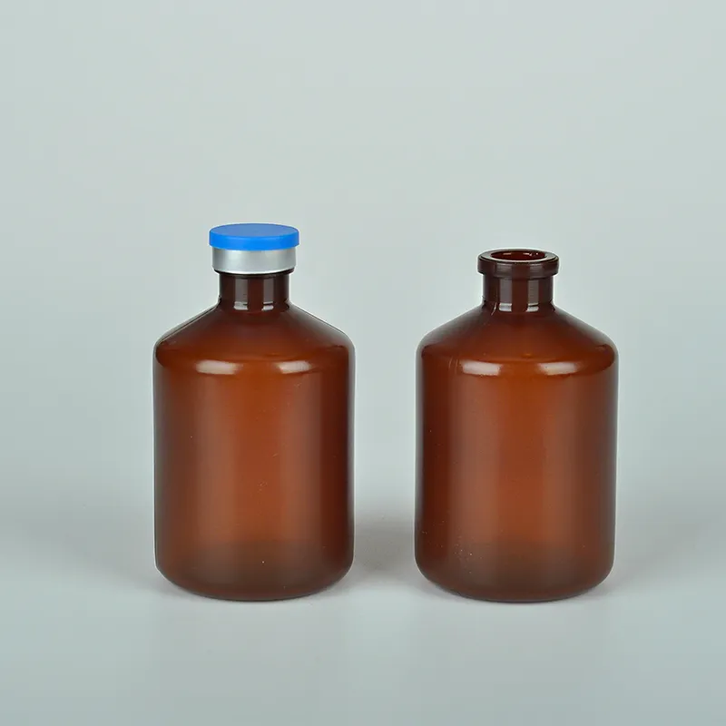
histology lab supplies
Understanding Histology Lab Supplies Essential Tools for Microscopic Examination
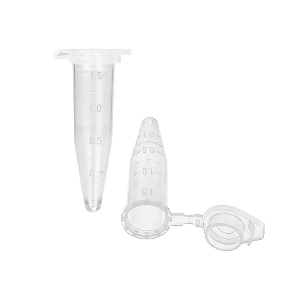
Histology, the study of the microscopic structure of tissues, is a crucial field in medical and biological research. It plays an indispensable role in diagnosing diseases, understanding tissue development, and advancing medical science. Central to this discipline are the histology lab supplies, which enable researchers and pathologists to prepare, examine, and analyze tissue samples efficiently. This article explores the essential supplies required in histology labs, their functions, and how they contribute to the accuracy and efficacy of histological studies.
Tissue Preservation and Processing
One of the primary steps in histology is the preservation of tissue samples. To prevent degradation and maintain the structural integrity of tissues, formalin (formaldehyde solution) is commonly used as a fixative. It cross-links proteins, thereby stabilizing cellular structures, which is critical for subsequent examinations. After fixation, tissues often undergo processing to remove water and infiltrate them with paraffin wax, a process facilitated by dehydrating agents such as ethanol and clearing agents like xylene.
In addition to fixatives, embedding materials are crucial. Paraffin wax is the most widely used embedding medium, as it provides support for thinly slicing the tissue samples into sections. The embedding process requires embedding cassettes to hold the tissues, along with molds for shaping the wax.
Sectioning and Staining
Once the tissues are embedded, the next step is sectioning. Microtomes or cryostats are the primary instruments utilized for slicing tissues into thin sections, typically between 3 to 10 micrometers thick. Microtomes are used for paraffin-embedded samples, while cryostats allow for rapid sectioning of frozen tissues.
After sectioning, staining is essential to enhance the visibility of tissue components under a microscope. Hematoxylin and eosin (H&E) staining is a foundational technique that differentiates cellular components hematoxylin stains nucleic acids blue, while eosin stains cytoplasmic structures pink. Beyond H&E, there are numerous special stains available, designed to highlight specific tissue types or components, such as Masson's trichrome for connective tissues or periodic acid-Schiff (PAS) for carbohydrates.
histology lab supplies
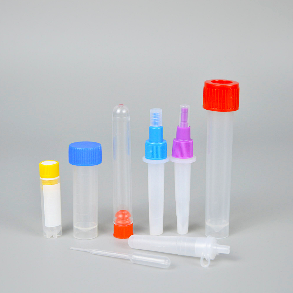
Microscopy
Observation of stained tissue sections is performed using microscopes. Light microscopes are most common, but in recent years, fluorescence and electron microscopes have gained prominence for examining cellular details with greater resolution. Each microscope type requires specific preparations in terms of glass slides and coverslips that protect the samples while allowing for clear visualization.
Safety and Quality Control
Safety is paramount in histology labs due to the use of hazardous chemicals and biological specimens. Personal protective equipment (PPE) such as gloves, lab coats, and goggles are essential for ensuring safety while handling toxic substances. Additionally, histology labs must adhere to stringent quality control measures, requiring supplies such as calibrated pipettes, analytical balances, and sterile containers to maintain accuracy and veracity in results.
Conclusion
Histology is an intricate field that forms the backbone of many medical diagnoses and research studies. The various supplies integral to histology labs—from fixatives and embedding materials to sectioning tools and staining reagents—each play a vital role in ensuring that tissues are preserved, prepared, and presented optimally for analysis. As technology advances, so too do the tools available for histology, embracing innovations that enhance precision, efficiency, and safety in laboratories worldwide.
In conclusion, understanding the diverse range of histology lab supplies underscores their significance in the rigorous processes of tissue examination. For specialists in the field, selecting high-quality materials and maintaining a keen awareness of safety protocols is essential for achieving accurate and meaningful results. As histology continues to evolve, so will the methodologies and tools that define it, ultimately enriching our understanding of the biological complexities within the tissues that sustain various life forms.
-
Aesthetic Makeup Spray Bottles | Fine Mist Empty RefillableNewsAug.19,2025
-
White Plastic Veterinary Vaccine Vials | Lab Liquid BottlesNewsAug.18,2025
-
Plastic Medicine Liquid Bottle: Secure Flip Top Drug VialsNewsAug.17,2025
-
Durable 250ml Blue Plastic Vaccine Vial for Lab & Vet UseNewsAug.16,2025
-
Sterile Virus Sample Tubes: Secure & Reliable Specimen CollectionNewsAug.15,2025
-
White 250ml Plastic Vaccine Vial for Lab & Vet MedicineNewsAug.14,2025


