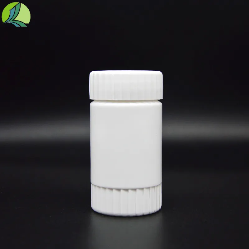
histology lab supplies
Histology Lab Supplies Essentials for Precise Tissue Analysis
Histology, the study of microscopic tissue structures, is fundamental to understanding the intricacies of biological systems. Within clinical and research settings, histology provides invaluable insights into disease pathology, developmental biology, and the effectiveness of therapeutic interventions. To conduct histological analyses accurately, the right lab supplies are indispensable. This article will explore the essential histology lab supplies, their importance, and how they contribute to precise tissue analysis.
1. Tissue Fixatives
Tissue fixation is the first crucial step in histological preparation. Fixatives serve to preserve tissue morphology and cellular structures, preventing degradation. Formalin, a common fixative, preserves proteins and nucleic acids while maintaining cellular architecture. Other fixatives, such as glutaraldehyde and Bouin’s solution, serve specific purposes depending on the type of study being conducted. The choice of fixative not only influences the integrity of the tissue samples but also affects subsequent staining protocols.
2. Paraffin Embedding Supplies
After fixation, the next step is embedding the tissue samples in paraffin wax, which provides support for thin sectioning. Paraffin embedding supplies include embedding molds, paraffin wax, and embedding stations. The quality of paraffin and its temperature play critical roles in ensuring that tissues are adequately infiltrated and preserved. Proper embedding also aids in obtaining uniform and thin sections for microscopic analysis.
3. Microtomes and Slide Preparation Tools
Microtomes are essential for slicing embedded tissue into thin sections, typically about 5-10 micrometers thick. Precision in this step is crucial, as uneven or thick sections can obstruct accurate microscopic evaluation. In addition to microtomes, supplies like glass slides, coverslips, and slide holders are essential for preparing and storing the tissue sections. These tools ensure that samples are accessible for staining and observation under a microscope.
4. Staining Reagents
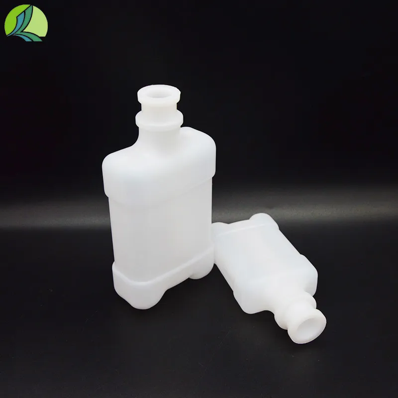
histology lab supplies

Staining is vital for visualizing specific cellular components within the tissue sections. Various dyes and reagents are employed depending on the structures of interest. Hematoxylin and eosin (H&E) staining is the most common method used in histology, providing a clear contrast between cellular components. Other specialized stains, like Masson's trichrome, periodic acid-Schiff (PAS) stain, and immunohistochemical stains, are used to highlight specific proteins or structures in the tissues. The quality of staining reagents significantly impacts the clarity and interpretability of histological slides.
5. Microscopes and Imaging Technology
After staining, the next essential supply is the microscope, a fundamental tool for histological analysis. Light microscopes are commonly used, but for more detailed studies, electron microscopes may be employed. Advances in imaging technology, including digital microscopy and imaging software, have revolutionized histological analysis, allowing for enhanced visualization and analysis of tissue structures. High-quality optics and imaging systems are crucial for obtaining accurate and reproducible results in histological evaluations.
6. Safety Equipment
Working in a histology lab often involves exposure to hazardous chemicals and biological materials. Therefore, safety equipment is paramount. Personal protective equipment (PPE), including lab coats, gloves, and goggles, is essential to ensure the safety of laboratory personnel. Additionally, fume hoods are necessary when working with volatile fixatives and reagents to minimize exposure to harmful fumes.
7. Storage Solutions
Proper storage solutions for reagents, samples, and equipment are critical to maintaining the integrity of histology lab supplies. Chemical preservatives for tissues, appropriate refrigeration for certain reagents, and organized storage for slides help prevent contamination and degradation. Ensuring proper labeling and organization in storage solutions allows for efficient workflow and easy retrieval of supplies when needed.
Conclusion
In conclusion, the field of histology relies heavily on a range of essential lab supplies that facilitate precise tissue analysis. From tissue fixation to microscopic imaging, each component plays a vital role in ensuring the accuracy and reliability of histological studies. As research and clinical practices continue to evolve, the demand for high-quality histology lab supplies will remain paramount for advancing our understanding of tissues and diseases. Investing in the right supplies not only enhances the quality of histological analysis but also contributes to better diagnostic and therapeutic outcomes in medicine and research.
-
Aesthetic Makeup Spray Bottles | Fine Mist Empty RefillableNewsAug.19,2025
-
White Plastic Veterinary Vaccine Vials | Lab Liquid BottlesNewsAug.18,2025
-
Plastic Medicine Liquid Bottle: Secure Flip Top Drug VialsNewsAug.17,2025
-
Durable 250ml Blue Plastic Vaccine Vial for Lab & Vet UseNewsAug.16,2025
-
Sterile Virus Sample Tubes: Secure & Reliable Specimen CollectionNewsAug.15,2025
-
White 250ml Plastic Vaccine Vial for Lab & Vet MedicineNewsAug.14,2025


